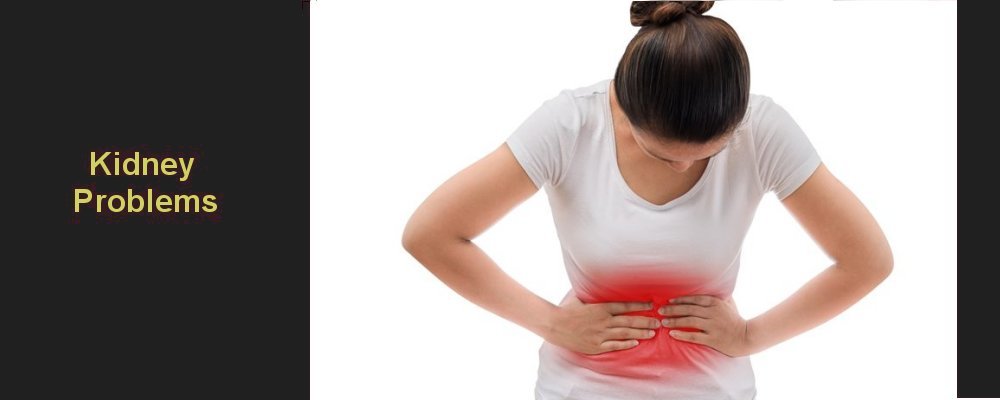
Kidney Problems |
||
|
||
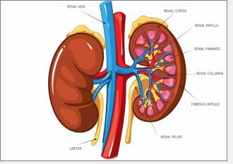
The kidneys remove waste and extra water from the blood to form urine. Urine flows from each kidney through a tube called the ureter to the bladder where it is stored until it is convenient to pass urine. From there, urine passes to the outside world through the prostate and urethra in men or the urethra alone in women. The kidneys are bean-shaped organs, and are about the size of a fist. They are located near the middle of the back, just below the rib cage. Each day, your kidneys process about 180 litres of blood to sift out about 1.5 litres of waste products and extra water. The waste and extra water become urine.
The wastes in your blood come from the normal breakdown and repair of bodily tissues and from food. Your body uses the food for energy and self-repair. After your body has taken what it needs from the food, waste is sent to the blood. If your kidneys did not remove these wastes, the wastes would build up in the blood and damage your body. Problems that can affect the kidneys includes:
Pelviureteric (PUJ) obstruction is the name given to a condition in which the flow of urine from the kidney is slowed down leading to pain in the back. Please click the links on the left to find out more about the condition and the operation to fix it.
To download information to print on PUJ obstruction and laparoscopic pyeloplasty, click here:
What is PUJ obstruction?
A blockage of the flow of urine from part of the kidney known as the renal pelvis to the ureter, which is the tube that carries urine onwards to the bladder.
What are alternative names for PUJ obstruction?
What causes PUJ obstruction?
There is usually an abnormality in the structure of the wall of the PUJ. This can exist from birth or develop later in life secondary to other causes such as stones or, very rarely, cancer. In about one in three cases to two in three cases, the PUJ passes over a blood vessel known as a ‘crossing vessel’ and this may cause the obstruction sometimes also. Even if PUJ obstruction is present in birth, symptoms may not occur until later in life.
What are the symptoms or features of PUJ obstruction?
In adolescents or adults, PUJ obstruction can cause pain on in the side of the back, and the pain can be worse after drinking. Other symptoms include
Occasionally, PUJ obstruction is eventually found after tests are made because blood is in the urine. None of these symptoms are specific for PUJ obstruction and the symptoms may be caused by other problems. Therefore, more action will be necessary to make a correct diagnosis.
What findings are made when an examination is made?
Rarely are any specific findings made. Occasionally, there may be swelling felt in the abdomen, or tenderness in the side of the back when the obstruction is significantly worse.
What tests can be done for this problem?
Tests are performed to show the changes characteristic of PUJ obstruction. These changes include an enlarged kidney and a delay in the passage of urine across the PUJ.
Why and when is treatment needed?
Reasons for treatment include
What treatments are available? click here
| ||
|
|
Summary |
|


