Kidney StoneTreatments |
||
|
Surgical Treatments
For larger stones that are unlikely to pass by itself or is causing too many problems then intervention may be required.
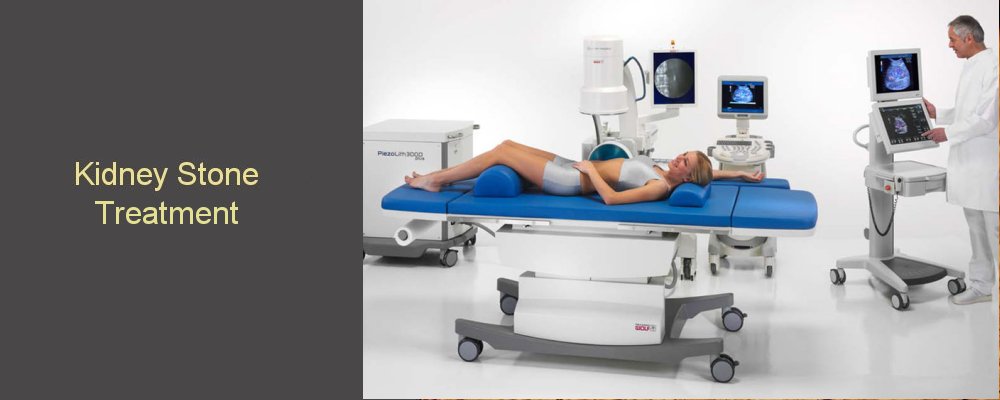
Extracorporeal Shock Wave Lithotripsy
ESWL is a non-invasive procedure. It involves treating a stone in the kidney or ureter without the need for a general anaesthetic. The patient lies on a bed then water filled cushion is then pressed up against the kidney and a shock wave is fired directed at the stone.
Extracorporeal shock wave lithotripsy (ESWL) uses sound waves or shock waves to break stones into small fragments that can pass spontaneously. It is performed usually as an outpatient procedure whilst awake or sometimes with sedation. Usually, you can go home immediately after, although it may need to be repeated What are the reasons for having ESWL? Usually, there is a stone present within the kidney or upper part of the ureter that will not pass by itself. The stone may sometimes cause pain or sometimes no pain at all, although it may impede the passage of urine from the kidney down to the bladder. Usually, the stone will be less than 2 cm in size, although sometimes larger. Stones present in the ureter may be treated where they are or sometimes pushed up into the kidney before treatment. To do this, a procedure under general anaesthetic would normally be required before the ESWL itself. What are the advantages of ESWL? There are several advantages of ESWL over other treatments for stones. What are the risks of ESWL? • About 1 in 10 people experience a problem. The main risks are: What are the alternatives to ESWL? Depending on the qualities of the stone and the individual with the stone, it may be possible to consider: • Watchful waiting (i.e. waiting for the stone to pass by itself)
ESWL may not be possible for patients with the following characteristics
• weight over 300 lbs (136 kg), You will usually be asked eat a light breakfast or lunch before the procedure. On the day of the procedure, you should wear comfortable clothes that are easy to remove, as you will have to change into a surgical gown. How is ESWL performed? When you arrive, you may need to change into different clothes. Sometimes, it is necessary to have another x-ray before treatment. After you enter the room with the ESWL machine, you will be asked to lie down on the treatment table. If the stone is in the kidney, you are usually asked to lie face down, but if the stone is in the ureters, you will probably be asked to lie on your back. The treatment usually lasts between 20 and 40 minutes; you will hear a “clicking” noise and feel something like a “flicking” on your back/front. Some people have said that it feels a bit like a small electric shock. The power or intensity may increase during the procedure so that the stone can be completely broken. It may feel uncomfortable but should be bearable and additional pain killers can be given. What to expect after ESWL? After passing urine, it should be possible to go home. Someone may need to drive you home. In some cases, it may be necessary to take antibiotics for a few days. You should large volumes of water to increase urine flow and help flush stone fragments through. You should be able to resume normal activities the day after treatment. You may see blood in your urine after the treatment. This is not important, unless the urine is completely opaque because of blood. A bruise may appear on the back where the treatment head was placed Small fragments of stone may pass giving pain sometimes as bad as renal colic. When should I contact a doctor? If you have the following, you should contact a doctor: How often does ESWL need to be repeated?
What happens if ESWL doesn't work?
One of the alternative options listed above may be needed i.e. either uretero-renoscopy or PCNL.
Ureteroscopy
Percutaneous Nephrolithotomy (PCNL)
PCNL is the surgical removal of stones from the kidney. This is reserved for larger stones or stones that cannot be treated with either ureteroscopy or shock wave lithotripsy. A general anaesthetic is required and a small (1cm) incision is made in the skin overlying the kidney. A telescope is then introduced through this incision into the kidney and the stone is broken and removed.
JJ stents There are different types of stents, and some of these differences allow a stent to provide different benefits depending on the situation . What's the reason for having a JJ stent? A JJ stent may be placed for several reasons. It allows urine to flow from the kidney to the bladder even when the ureter is blocked for one reason or another. This way, the kidney keeps working and is not damaged by being obstructed and avoids the severe pain that can occur when a kidney does not drain properly. The chance of an infection is also reduced significantly. A stent protects the ureter and allows the ureter to heal even when damaged. If a stent is not placed and the ureter is hurt in some way or other, it can become too narrow when it heals forming what is called a stricture. Having a stent can prevent that from happening and makes it more likely that the ureter will work well afterwards. Sometimes, a stent is placed because it makes a narrow ureter wider over a period of time. This can be important when access through the ureter is needed to pass instruments or remove stones. This would typically occur when an attempt to go up the ureter to get a stone has failed because it was too narrow. Inserting a stent makes it more likely that later attempts to get up the ureter will be successful. What are the disadvantages of having a JJ stent? It is not possible to predict who will or will not have side-effects with a stent. Some people tolerate stents without problems. Others find they have problems described below. Such problems may be present only at the beginning of having a stent and resolve over a few days or weeks. Other people may find their symptoms persist through out the period of the stent being present. Stents can cause blood to appear in the urine at various times. Usually, physical activity of one kind or other results in movement of the stent inside the body. This can give rise to blood in the urine. Pain may be felt in the back (loin), bladder area, groin, penis in men or urethra in women, and sometimes the testicles. The discomfort or pain may be more noticeable after physical activities and after passing urine. The stent can cause irritation of the bladder and so make it necessary to pass urine more frequently including the need to get up at night to pass urine. These symptoms can sometimes be improved by medication. Rarely, a stent may cause a woman to leak urine. Once the stent has been removed, these side-effects go away.
What problems can arise with a stent? Sometimes, stents can become calcified and develop a coating similar to stones. Stents can also move out of position. When this happens, the stent usually moves into the bladder causing a deterioration in bladder symptoms i.e. going to the toilet to pass urine more frequently, discomfort in the area of the bladder and perhaps blood in the urine. How does a stent interfere with daily life? You can still go to work and play sports when you have a stent in place. However, you may feel more tired and experience discomfort during the day limiting your performance. In addition, you may need to visit a toilet more frequently and so need convenient access to a toilet. Travel is possible, although medical attention may be required rarely. As stents can have side-effects, your ability to enjoy yourself may be limited as a result. What additional care is necessary when a stent is in place? Drink at least 1½ to 2 litres (approximately four pints) of fluids a day When might it be necessary to call a doctor?
You should contact a doctor if How is a stent inserted? A stent is inserted usually under a general anaesthetic often in combination with another procedure depending on the reason for the stent. A telescope called a cystoscope is passed through the urethra (water pipe) and into the bladder. The stent is passed through the cystoscope and into the ureter. The position of the stent is checked with x-rays. What alternatives are there to a JJ stent? Sometimes, it may be reasonable not to leave a JJ stent if obstruction is likely to be transient. This may be risky and depends on the circumstances. If several procedures have been performed, there is often swelling making obstruction and pain a distinct possibility. Occasionally, it may be possible to place a tube internally draining the kidney that comes out through the urethra (water pipe). This can simply be removed by pulling it out without needing a further procedure of any kind. The disadvantage is that it can remain for only a day or so. Another alternative is to have tube placed directly through the skin and into the kidney. This is called a 'nephrostomy'. This is placed under guidance by ultrasound and the kidney has to be distended to get into the correct place without difficulty. As this is outside the body, it is slightly more inconvenient and can sometimes get pulled out by accident. Its advantage is that it usually drains better than a JJ stent which can be important if there is infection with obstruction of the kidney ('pyonephrosis'). How is a stent removed? A JJ stent is removed with a cystoscope performed under local or general anaesthetic. A special flexible telescope is passed through the urethra. The stent is picked up and removed. Please see flexible cystoscopy.
|
|
|
||


